
To reference this electronic educational resource according to the APA style for Web references, use:
Liem EB (2006): Bullard Laryngoscope Intubation. Retrieved <insert date of retrieval here>, from University of Florida Department of Anesthesiology, Center for Simulation, Advanced Learning and Technology, Virtual Anesthesia Machine Web site: http://vam.anest.ufl.edu/airwaydevice/bullard/index.html
Keywords: Bullard laryngoscope, Intubation, difficult airway, video-intubation
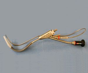
Bullard Laryngoscope (ACMI)
See Bullard videos in the video library
Introduction
The Bullard laryngoscope is one of several different types of rigid fiberoptic laryngoscopes that are available (1). The rigid fiberoptic laryngoscopes aids with visualization of the glottic opening even when there is an inability to align the oral, pharyngeal, and laryngeal axes. Compared to conventional direct laryngoscopy the rigd fiberoptic laryngoscopes only require minimal head manipulation and positioning (3, 4). In addition, the Bullard laryngoscope can be used in patients with a mouth opening of as little as 6 mm.
The Bullard laryngoscope can assist in both oral and nasal intubations (5, 6). For nasal intubations, the Bullard laryngoscope is introduced into the mouth for visualization of the vocal cords but the ETT is not loaded onto the dedicated stylet. With experience the Bullard laryngoscope allows for rapid visualization of the larynx and can be used for rapid sequence intubation. Awake intubation is possible but requires good patient preparation and considerable skill (2,7). It can be used in the pediatric population, since a pediatric sized laryngoscope and stylet are available.
These above mentioned features make the Bullard laryngoscope a good option for certain scenarios where conventional direct laryngscopy might prove to be very difficult : patients with anterior larynx, unstable cervical spine fracture, upper body burns, trauma, patients with TMJ immobility and micrognathia (e.g. Pierre Robin syndrome).
We will describe the preparation and sequence of events when using the Bullard laryngoscope with its dedicated stylet for oral intubation in a patient induced with general anesthesia (8,9).
Preparation
The Bullard laryngoscope comes in both an adult and pediatric size. A stylet should be used to guide the ETT during intubation. This can be the dedicated unmalleable wire stylet, which has to be attached to the laryngsocope, or an independent malleable stylet. Alternatively, there is also a multifunctional stylet available which has the same shape as the old stylet and attaches in the same way. The multifunctional stylet is hollow which allows for the passage of an airway catheter, suctioning under direct vision, or oxygen administration. A fiberoptic lightsource and the blade/tip extender should be available. The choice between the adult or pediatric size laryngoscope is a function of both the height of the patient ( < 5 ft pediatric, > 5ft adult, > 6ft adult with tip extender), and the minimal size of the ETT that can be fitted onto the adult stylet. Some find the tip extender useful in patients as small as 5'4", depending on their mandibular angle to thyroid cartilage distance.
- Place tip/blade extender onto laryngoscope blade - make sure the tip extender is securely snapped into place (the extender is only used for the adult bullard).
- Select the appropriate size ETT for the patient. Temporarily remove the ETT connector when using smaller sizes, as this will facilitate advancing the ETT over the stylet during intubation. Apply a water-based lubricant (e.g. Lidocaine jelly, K-Y jelly, Hurricaine spray,) then insert stylet into ETT until tip extends out of the regular opening.
- When using the unmalleable wire stylet, it should be attached to the laryngoscope handle. When looking through the optical eyepiece, the stylet should be seen through the lens but not the ETT. It should be withdrawn until it is out of view if the ETT is also seen.
- If needed, apply water based lubricant to blade section to facilitate insertion of the blade.
- Test fiberoptic lightsource and snap onto ETT/stylet/laryngoscope assembly. A guard for the fiberoptic lightsource can also be used; snap it into place by depressing the knob located directly behind the fiberoptic source attachment.
- Make optical element non-wettable by applying silicon oil (this will have better adherence to the lens than anti-fog solution.)
- Consider giving the patient an anti-sialogogue.
 |
 |
||||||||||||||
 |
 |
||||||||||||||
 |
|||||||||||||||
 |
|||||||||||||||
 |
|||||||||||||||
 |
 |
||||||||||||||
 |
|||||||||||||||
 |
|||||||||||||||
 |
|||||||||||||||
Oral Intubation: A Step by Step Guide
- Induce patient as if for regular intubation; apply cricoid pressure if rapid sequence intubation is desired.
- Patient position should be undisturbed or neutral, not "sniffing".
- Position yourself as for regular direct laryngoscopy and position the scope axis parallel to patient axis pointing caudad (see below for illustration.)
- Introduce the scope tip between the teeth, into the pharynx. Try to stay as close to midline as possible.
- The ETT is held firmly in the "nook" of the Bullard as the laryngoscope/ETT assembly is advanced as a single unit into the mouth and pharynx.
- Advance the laryngoscope/ETT assembly over the tongue using the left hand, sweep to a position perpendicular to the patient axis minimally distorting the anatomy.
- Elevate the bullard handle straight up, the blade tip should retract the epiglottis and you should be able to visualize the glottic opening. It should be noted that this is NOT a passive technique where the scope is simply advanced into the pharynx for visualization. SUSPENSION laryngoscopy is required to establish the viewing space necessary to identify the epiglottis and ultimately the larynx. When performing the suspension laryngoscopy, the right hand can be placed on the laryngoscope for stabilization. Alternatively, some prefer to use the right hand to grab the mandible (thumb in the mouth, fingers on mentum) and elevate it so that less traction can be applied to the Bullard laryngoscope, thereby making maneuverability easier
- Advance the ETT over the stylet using the right hand.
- Detach the stylet from the laryngoscope and remove the larygnoscope in the opposite motion as during intubation.
- Withdraw the stylet and confirm correct position as for regular intubation.
Troubleshooting Tips
Cannot see vocal cords
Usually this means one is not exactly midline. Because the view through the Bullardscope is less panoramic than regular intubation, it is harder to correct during laryngoscopy if you are not entering the mouth perfectly in the midline. Step back and determine if you are perfectly midline with the laryngoscope. Feel the trachea / thyroid cartilage to determine the midline again, then re-advance the laryngoscope.
Another approach would be to elevate the jaw with the right hand while looking through the eyepiece at the same time. Then attempt to visualize the vocal cords; the decreased amount of pressure on the blade will make it easier to maneuver the blade.
Difficulty advancing the ETT with the vocal cords visualized
The laryngoscope is advanced too far and advancement of the stylet is obstructed by the arytenoids. Usually this means that you are too close to the larynx. Remember that the viewing lens is to the side of the ETT; as you advance it off the stylet, it will travel from its coaxial position to a position directly in line with the view from the lens. Withdraw the laryngoscope slightly in the horizontal plane and re-advance the ETT.
Webauthor: E.B. Liem
Consultants:
D.G. Bjoraker
R. Bullard (inventor of the Bullard Laryngoscope)
D.Gravenstein
Contributors: A.J. Deckinga
- Bjoraker-DG. The Bullard intubating laryngoscopes. Anesth Rev 17(5): 64-70, 1990.
- Cohn-AL, Zornow-MH. Awake endotracheal intubation in patients with cervical spine disease: A comparison of the Bullard laryngoscope and the fiberoptic bronchoscope. Anesth Analg 81:1283-1286, 1995.
- Hastings-RH, Vigil-AC, Hanna-R, Yang-BY, Sartoris-DJ. Cervical spine movement during laryngoscopy with the Bullard, Macintosh, and Miller laryngoscopes. Anesthesiology 82:859-869, 1995.
- Watts-ADJ, Gelb-AW, Bach-DB, Pelz-DM. Comparison of the Bullard and Macintosh laryngoscopes for endotracheal intubation of patients with a potential cervical spine injury. Anesthesiology 87:1335-1342, 1997.
- Shigematsu-T, Miyazawa-N, Kobayashi-M, Yorozu-T, Toyoda-Y, Morisaki-H. Nasal intubation with Bullard laryngoscope: A useful approach for difficult airways. Anesth Analg 79:132-135, 1994.
- Brown-RE jr, Vollers-JM, Rader-GR, Schmitz-ML. Nasotracheal intubation in a child with Treacher-Collins syndrome using the Bullard intubating laryngoscope. J Clin Anesth 5:492-493, 1993.
- Cohn-AL, MCGraw-SR, King-WH. Awake intubation of the adult trachea using the Bullard laryngoscope. Can-J-Anaesth 42:246-248, 1995.
- Cooper-SD, Benumof-JL, Ozaki-GT. Evaluation of the Bullard laryngoscope using the new intubating stylet: Comparison with conventional laryngoscopy. Anesth Analg 79:965-970, 1994.
- Cohn-AI, Isaac-P, Ramakrishnan-U, Harbourne-K. Bullard laryngoscope: Preliminary experience with the new multifunctional stylet. J Clin Anesth 10:681-683, 1998.