
To reference this electronic educational resource according to the APA style for Web references, use:
Liem EB (2006): Upsher Laryngoscope Intubation. Retrieved <insert date of retrieval here>, from University of Florida Department of Anesthesiology, Center for Simulation, Advanced Learning and Technology, Virtual Anesthesia Machine Web site: http://vam.anest.ufl.edu/airwaydevice/upsher/index.html
Keywords: Upsher Laryngoscope, Intubation, difficult airway, airway management, video-intubation
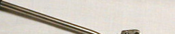
Upsherscope (Mercury Medical)
See Upsher videos in the video library
Introduction
The Upsher laryngoscope (Upsherscope) is one of several different types of rigid fiberoptic laryngoscopes that are available. These rigid fiberoptic laryngoscopes aid with visualization of the glottic opening even when there is an inability to align the oral, pharyngeal, and laryngeal axes. Compared to conventional direct laryngoscopy, the rigid fiberoptic laryngoscopes require minimal head manipulation and positioning. The Upsherscope requires a minimal mouth opening of 15 mm, less than required for conventional direct laryngoscopy.
Use of the Upsherscope has been described for only oral intubations. With experience it allows for rapid visualization of the larynx and can be used for rapid sequence intubation. Awake intubation is possible but requires good patient preparation and considerable skill (2). Only an adult version of the Uspherscope is available; there is no pediatric version.
These above mentioned features make the Upsherscope a good option for
certain scenarios where conventional direct laryngscopy might prove
to be very difficult: patients with anterior larynx, unstable cervical
spine fracture, upper body burns, trauma, TMJ immobility, and micrognathia.
However, compared to conventional direct laryngoscopy, the Upsherscope
can be technically more difficult to use: and a published prospective
randomized controlled clinical trial (1) actually showed a higher failure
compared to direct laryngoscopy when used in the general population
(not selected for difficult airways.) We will describe the preparation
and sequence of events when using the Upsherscope for oral intubation
in a patient induced with general anesthesia.
Preparation
A fiberoptic lightsource should be available. The Upsher laryngoscope will fit an 7.0, 7.5 or 8.0 ETT. Smaller ETTs are not optimally guided. Larger ETTs will not slide very well or not fit at all.
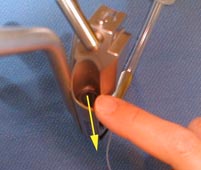
- Remove the ETT connector. Apply a water-based lubricant (e.g. Lidocaine jelly, K-Y jelly, Hurricaine spray) to the outside of the ETT. Place the ETT in the tube guiding channel with the tip of the ETT protruding approximately 1 cm beyond the channel and with the ETT rotated 90 degrees anti-clockwise (bevel posterior).
- Focus the scope and confirm by looking through the eyepiece that the tip of ETT is visible but without having the ETT obscuring the view.
- Test fiberoptic lightsource, snap and lock it on the Upsherscope.
- Make optical element non-wettable by applying silicon oil (this will have better adherence to the lens than anti-fog solution.)
- Consider giving the patient an anti-sialogogue.
 |
 |
 |
|||||||||||||||
 |
|||||||||||||||||
 |
 |
||||||||||||||||
 |
|||||||||||||||||
 |
|||||||||||||||||
 |
|||||||||||||||||
 |
|||||||||||||||||
Oral Intubation: A Step by Step Guide
- Induce patient as if for regular intubation; apply cricoid pressure if rapid sequence intubation is desired.
- Patient position should be undisturbed or neutral, not "sniffing". Some authors even propose a slight flexion of the head and neck to increase the chance of the scope picking up the epiglottis.
- Position yourself as for regular direct laryngoscopy and position the scope axis parallel to patient axis pointing caudad (see the animation in the Bullard laryngoscopy section.)
- Introduce the scope tip between the teeth, into the pharynx. Try to stay as close to midline as possible.
- Advance the laryngoscope/ETT assembly over the tongue, sweep to a position perpendicular to the patient axis minimally distorting the anatomy. The aim is to pick up the epiglottis by a sweeping motion.
- Elevate the Upsherscope handle straight up. The blade tip should
retract the epiglottis and you should be able to visualize the glottic
opening. It should be noted that this is NOT a passive technique
where the scope is simply advanced into the pharynx for visualization.
SUSPENSION laryngoscopy is required to establish the viewing
space necessary to identify the epiglottis and ultimately the larynx.
When performing the suspension laryngoscopy, the right hand can be
placed on the laryngoscope for stabilization. Alternatively, some
prefer to use the right hand to grab the mandible (thumb in the mouth,
fingers on mentum) and elevate it so that less traction can be applied
to the Upsher laryngoscope, thereby making maneuverability easier.
Troubleshooting Tips
Cannot see vocal cords
Usually this means one is not exactly midline. Because the view through the Upsherscope is less panoramic than during regular intubation, it is harder to correct during laryngoscopy if you are not entering the mouth perfectly in the midline. Step back and determine if you are perfectly midline with the laryngoscope. Feel the trachea or thyroid cartilage to determine the midline again, then re-advance the laryngoscope.
Another approach to the above problem would be to elevate the jaw with the right hand while looking through the eyepiece at the same time. Then attempt to visualize the vocal cords, the decreased amount of pressure on the blade will make it easier to maneuver the blade.
Another reason for difficult visualization can be due to the inability to pick up the epiglottis (1). Because of the upward curve of the tip of the Upsherscope, the blade tip is often not seen during intubation which can make it difficult to position the tip underneath the epiglottis. Picking up the epiglottis is often a blind maneuver. As mentioned previously, increasing flexion of the head and neck could facilitate picking up the epiglottis.
Difficulty advancing the ETT through the vocal cords
The design of the Upsherscope is such that the tip of the ETT tends to be directed posteriorly and to the right of the visual field by the tube guiding channel of the Upsherscope. The main difficulty in advancing the ETT is usually due to the fact that it will tend to advance off to the right and posterior (3). Some authors have suggested loading the ETT 90 degrees anti-clockwise in the tube guiding channel as this will direct the ETT more towards midline. Even higher succes rated have been achieved by using the more maneuverable gum elastic bougie inside the ETT as a guide (2, 3). The tip of the gum elastic bougie is directed medial and the bougie is advanced through the vocal cords first.
Webauthor: E.B.Liem
Consultant: D.Gravenstein
- Fridrich-P, Frass-M, Krenn-CG, Weinstable-C, Benumof-JL, Krafft-P. The Upsherscope in routine and difficult airway management: A randomized, controlled clinical trial. Anesth Analg 85: 1377-1381, 1997.
- Pearce-AC, Shaw-S, Macklin-S. Evaluation of the Upsherscope: A new rigid fiberscope. Anaesthesia 51: 561-564, 1996.
- Yeo-V, Chung-DC, Hin-LY. A bougie improves the utility of the UpsherScope. J Clin Anesth. 1999 Sep; 11(6):471-6.